You will be discharged from the hospital the same day. You will wear an eye bandage and a protective cap to prevent eye injuries. These can be removed after a few hours.
Visual acuity returns to normal a few days after surgery, but this depends on the patient and the severity of the cataract. The more advanced the cataract, the longer the recovery may take.
A treatment is prescribed for 6 weeks, consisting of an antibiotic drop and an anti-inflammatory to prevent possible inflammation.
There may be some symptoms after the operation, but these are not alarming:
The eyes may be red and feel gritty. This is normal and is due to healing, it may take a few days.
Blurred vision on the day of surgery and the following day is normal.
Double vision may occur due to the anesthesia, but this is short-lived.
You can resume your daily activities without problems, but be careful with sports. The eye remains fragile, even when the vision is clear again. For sports that cause shocks, it is recommended to wait 3 weeks. You can wash your hair, but avoid contact of the eye with water. You can keep your old glasses or replace them with glasses with neutral lenses if they cause discomfort.
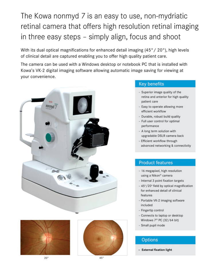
An investment in precision and care: the Jodoigne Eye Center, our sister practice within the same care chain, has equipped itself with the KOWA 7 retinograph (Made in USA).
To ensure even more accurate monitoring of our patients, the center recently invested in a KOWA 7 retinograph — a high-tech device from the United States.
With an impressive image resolution of 16 megapixels, this device takes razor-sharp images of the retina, allowing even the smallest invisible structures to be visualized in detail.
This technology allows us to closely monitor the course of important retinal structures such as the optic nerve, the macula and the retinal blood vessels. This is essential in the diagnosis and monitoring of conditions such as glaucoma, age-related macular degeneration (AMD), thrombosis, hypertension and of course diabetes.
A valuable step forward for personalized and quality eye care.
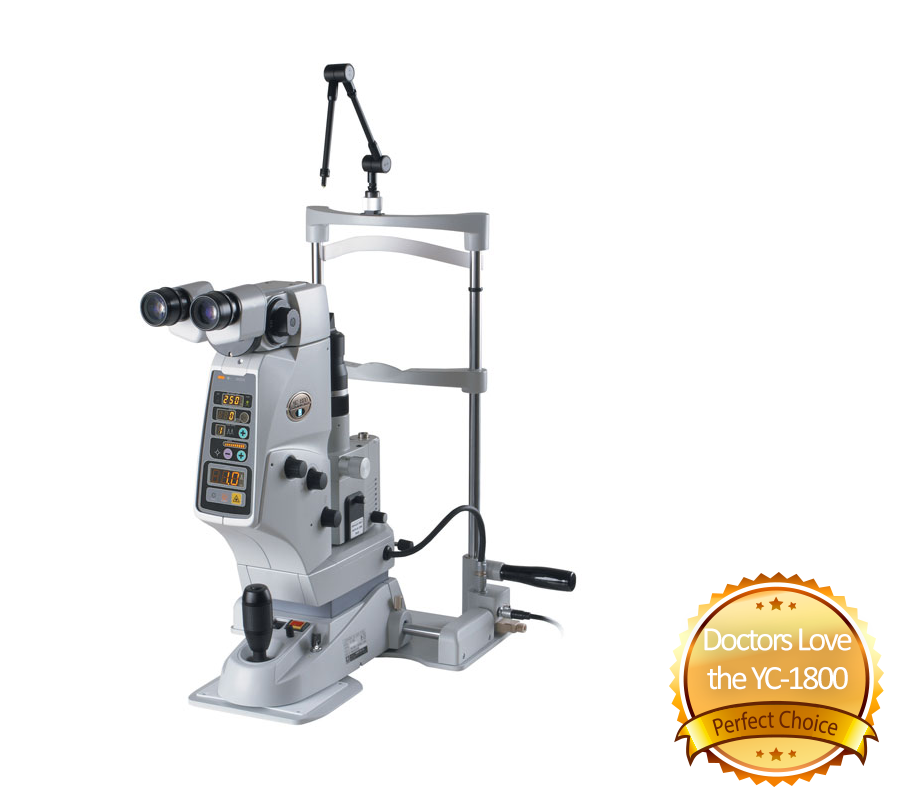
LASER YAG permet le traitement de : – la cataracte secondaire par une capsulotomie – prévenir le glaucome aiguë par fermeture de l’angle par une iridotomie
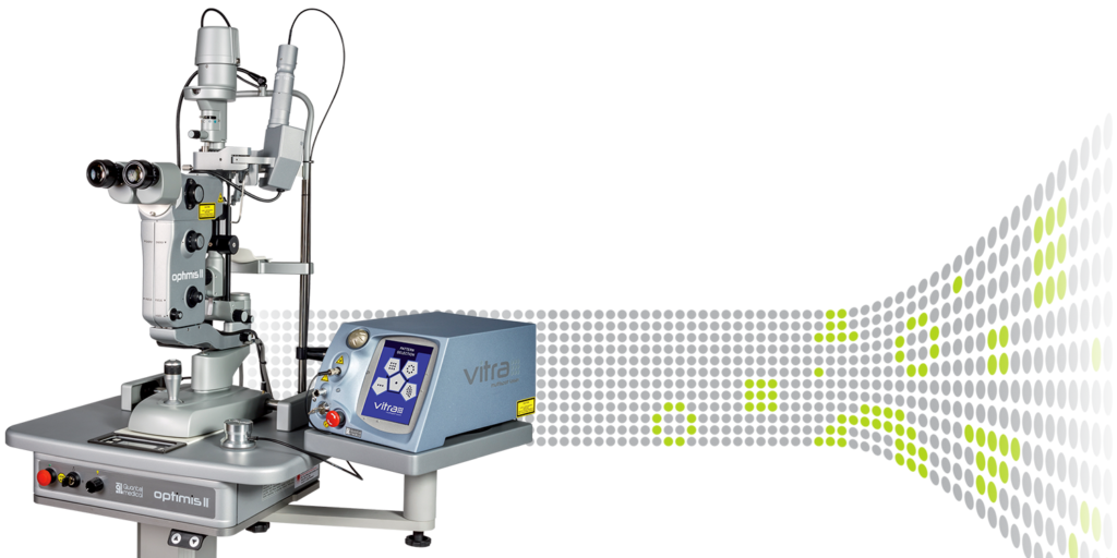
LASER ARGON Le traitement des : Maladie rétiniennes (Diabète, Déchirure rétinienne, post Thrombose)
Photographic Splintern Lamp A splinter lamp is a device used to take detailed images of the eyes, such as the eyelids, cornea, lens, and retina, to help diagnose eye diseases and other conditions. Adding “photographic” refers to the ability to capture images of the eyes for further analysis.
Retinograph + Autofluorescence These technologies are used to examine and analyze the health of the retina, especially for detecting eye diseases such as macular degeneration or diabetic retinopathy.
OCT (Optical Coherence Tomography) OCT is an advanced imaging technique used to obtain detailed cross-sectional images of the retina and other structures in the eye. It is essential for the diagnosis and follow-up of conditions such as glaucoma, macular degeneration and diabetic retinopathy.
OCT (Optical Coherence Tomography) OCT is an advanced imaging technique used to obtain detailed cross-sectional images of the retina and other structures in the eye. It is essential for the diagnosis and follow-up of conditions such as glaucoma, macular degeneration and diabetic retinopathy.
Visual Field The visual field test is used to measure a patient's peripheral (side) vision capacity and to detect abnormalities that may indicate conditions such as glaucoma, neurological problems or retinal disorders.
Goldmann visual field examination This is a specific type of visual field examination in which manual perimetry is performed. It is mainly used in patients who have difficulty with automatic perimetry or when a more detailed analysis of the peripheral visual field is required, such as in neurological disorders or advanced glaucoma.
Prescription Meter It is an instrument used to measure the strength of spectacle lenses (and sometimes contact lenses), including the strength of the spherical and cylindrical correction, the axis and possibly the prism.Automatic refractor quickly and accurately measures the basic strength of your eyes (nearsightedness, farsightedness and astigmatism), often as the first step in an eye examination.Autorefractor keratometer, tonometer and pachymeter
Autorefractor-keratometer: A device that automatically measures the refraction (strength of the eye) and the curvature of the cornea (keratometry).
Tonometer: An instrument that measures eye pressure, essential for the detection of glaucoma.
Pachymeter: A device that measures the thickness of the cornea, important in evaluating eye pressure and prior to certain surgical procedures such as LASIK.
Autorefractor keratometer, tonometer and pachymeter