
News
-
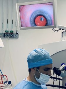
Experience cataract surgery step by step
Practical example – Cataract surgery on Mrs. H. (55 years old)
Date of surgery: Monday, February 22, 2021
Situation before the procedure
- Visibility: only shadows visible at 3 meters distance
- Diagnosis: very dense cataract, which severely limited daily functioning
Course of the surgery
- The natural cloudy lens was removed through a micro-incision of only 2 mm
- A foldable implant lens was carefully injected
- The artificial lens unfolded by itself and fell into place perfectly in the lens capsule
- The positioning was optimal and the procedure went smoothly
After a few minutes
- The eye structures were stable
- The clarity of the artificial lens was immediately visible
- The patient was able to perceive light and contours better shortly after the operation
🌟 This example illustrates how even in advanced cataracts, safe, precise and effective intervention is possible with modern microsurgery.
Would you like to know whether cataract surgery is also a solution for you?
📅 Make an appointment for a consultation.Before the procedure: Dense cataract
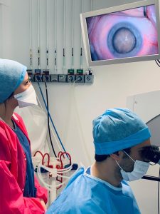
Insertion of the folded implant lens through a 2 mm micro-incision
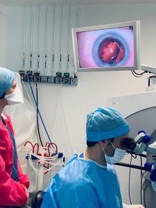
Good positioning of the implant lens
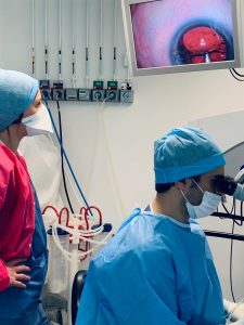
Image a few minutes after the procedure
-
Interview with a Patient (Video)
In this video, one of our patients shares her personal experience with the eye treatment she underwent at our center.
Watch the interview and discover how our expert care and advanced technology have made a difference in our patients' lives.
-
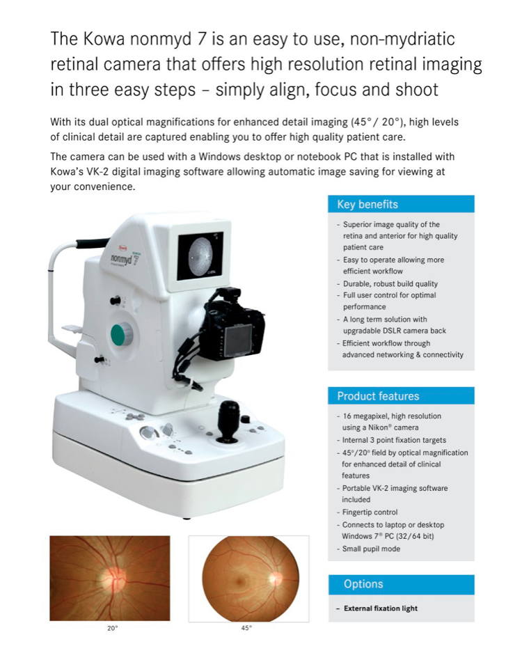
The Jodoigne Eye Center now has a new device for retinal examination
An investment in precision and care: the Jodoigne Eye Center, our sister practice within the same care chain, has equipped itself with the KOWA 7 retinograph (Made in USA).
To ensure even more accurate monitoring of our patients, the center recently invested in a KOWA 7 retinograph — a high-tech device from the United States.
With an impressive image resolution of 16 megapixels, this device takes razor-sharp images of the retina, allowing even the smallest invisible structures to be visualized in detail.
This technology allows us to closely monitor the course of important retinal structures such as the optic nerve, the macula and the retinal blood vessels. This is essential in the diagnosis and monitoring of conditions such as glaucoma, age-related macular degeneration (AMD), thrombosis, hypertension and of course diabetes.
A valuable step forward for personalized and quality eye care.
-
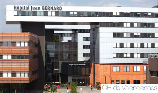
Samenwerking met het Oogcentrum van het Jean Bernard Ziekenhuis in Valenciennes.
De praktijk draagt bij aan de zorg en continuïteit van de behandeling van patiënten die worden gevolgd tussen het ziekenhuis van Valenciennes en Maubeuge.
Het lasercentrum van de kliniek du Parc in Maubeuge is het enige volledige behandelplatform voor refractieve chirurgie tussen Maubeuge, Valenciennes en de rest van het Avesnois-gebied.
Deze investering in de kliniek du Parc komt ter aanvulling op een globale en complementaire zorgaanbieding, samen met het ziekenhuis Jean Bernard, dat gespecialiseerd is in de behandeling van ernstige aandoeningen, met name van het netvlies.
Lasertechnologie voor het corrigeren van visuele afwijkingen wordt niet vaak aangeboden binnen Franse ziekenhuizen, met uitzondering van het CHRU, dat beschikt over een Excimer-laser.
De belangrijkste reden hiervoor is dat deze innovatieve technologie op dit moment niet wordt vergoed door de FRanse sociale zekerheid.
Sommige Franse ziektekostenverzekeraars vergoeden een deel van de kosten; het is raadzaam om bij uw verzekeraar navraag te doen.