This technique is still under development, and we will only offer it to our patients once the manufacturers have made significant progress.
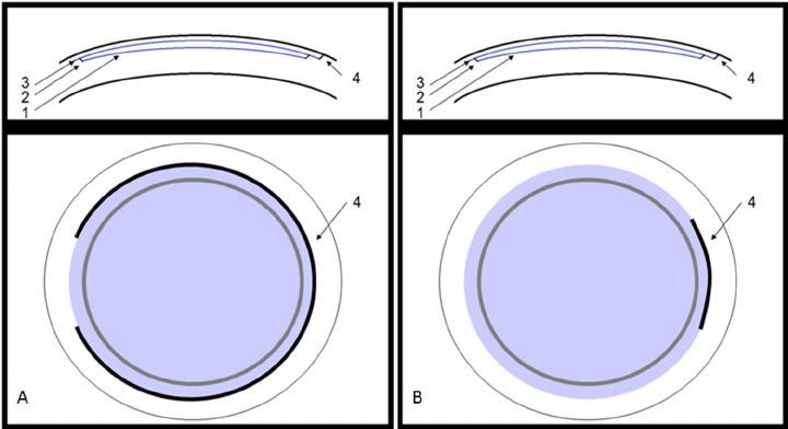
KLEX (Keratorefractive Lenticule EXtraction) is an advanced laser vision correction technique used to treat myopia (nearsightedness) and astigmatism, without creating a corneal flap. It is part of the same family as SMILE, SILK, CLEAR, and SmartSight.
Procedure Steps
- The femtosecond laser creates a small lenticule (disc) in the cornea.
- The surgeon manually removes this lenticule, reshaping the cornea to correct the refractive error.

Platforms and Technologies
- Zeiss – SMILE (Visumax 500 of 800)
- Johnson & Johnson – SILK (Elita-laser)
- Ziemer – CLEAR (Z8-laser)
- Schwind – SmartSight (Atos-laser)
All these techniques share the same principle: a lenticule is created and manually extracted to reshape the cornea.
Technical and Clinical Considerations
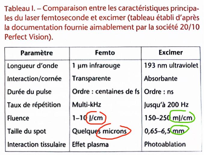
The KLEX technique does not use an excimer laser, making it less costly than LASIK.
However, the excimer laser offers submicron precision and a smooth, continuous ablation, allowing for more accurate and extensive corrections.
The femtosecond laser, on the other hand, works with 5–10 micron pulses, in a dot pattern, which is less refined.
Despite its image as a “less invasive” technique, KLEX remains more manual than LASIK: the surgeon must mechanically loosen the surfaces of the lenticule with a spatula and remove it through a small side incision.
To avoid tearing the lenticule, the edge must be at least 10 microns thick, meaning that about 15 microns of additional tissue are removed – with no optical benefit, just for the extraction.
Advantages and Limitations
The theoretical benefits (less corneal cutting, better nerve preservation, less dry eyes) have not yet been convincingly proven.
Potential drawbacks include:
- manual centration (no automated eye tracker)
- meticulous dissection to avoid residual fragments or folds
- increased risk of decentration
Postoperative Recovery
Temporary blurring of vision is common in the first few weeks or months due to the mechanical stress of the dissection.
The final results are comparable to LASIK, but without the typical “WOW effect” (sharp vision the very next day).
Biomechanical Aspects
The often-cited greater corneal stability with KLEX compared to LASIK or PRK (especially with thin corneas) remains theoretical and has not been confirmed by clinical studies.
A cornea that is at risk for LASIK is also at risk for SMILE or KLEX.
Complications in SMILE and LASIK
Intraoperative (during surgery)
- Loss of suction
- Black spots
- Abrasions
- Epithelial incision tear
Specific to SMILE / KLEX:
- Difficult dissection (OBL – opaque bubble layer)
- Lenticule tear
- Lenticule remnants
- Cap perforation
- Incomplete lenticule extraction
Postoperative (after surgery)
- Corneal haze
- Irregular astigmatism
- Ectasia
- Interface infection
- Interface inflammation (keratitis)
- Loss of best-corrected visual acuity (BCVA)
- Blurred or “foggy” vision
- “Rainbow glare”
- Cap displacement or folds (specific to LASIK)
